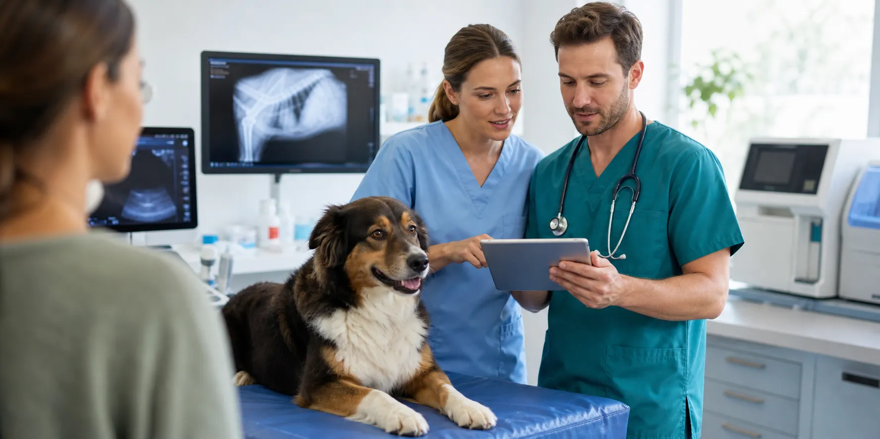
When the physical exam raises questions, imaging provides answers. Digital X-rays show us fractures, arthritis, swallowed objects, bladder stones, heart size, and lung disease within minutes; ultrasound adds a real-time look inside organs, revealing masses, fluid, and structural changes X-rays can't show.
For complex cases, CT (computed tomography) scanning provides detailed cross-sectional views of bone, soft tissue, the chest, and the skull — invaluable for surgical planning, nasal and ear disease, and subtle fractures.
Most studies are done during your visit with gentle positioning — sedation only when it makes your dog more comfortable. Complicated cases are sent for board-certified radiologist review.
Imaging services
- Digital X-rays with rapid results
- Abdominal ultrasound
- CT (computed tomography) scans
- Orthopedic and spinal imaging
- Foreign body and obstruction evaluation
- Cardiac and pre-surgical screening
- Radiologist consultation on complex studies
What to expect
Most studies happen during your visit: digital X-rays take minutes with gentle positioning, ultrasound adds a real-time look inside organs, and findings are reviewed with you the same day. Sedation only when it makes your dog more comfortable; radiologist review for the complicated cases.
Ultrasound
- Non-invasive, real-time imaging of the abdomen and heart
- No anesthesia needed for most patients
- Helps evaluate organs X-rays can't fully show
- Results reviewed with you the same visit whenever possible
Imaging guides
Dog Diagnostic Imaging guides & handouts
Take-home guides from our resource library related to this service.
Frequently asked questions
My dog swallowed something. Will an X-ray show it?
Bones, metal, and rocks show clearly; fabric, plastic, and rubber often don't show directly — instead we read the gas patterns they create, sometimes adding contrast or ultrasound. Bring the packaging or a matching object if you have it; it genuinely helps.
What's the difference between an X-ray and an ultrasound?
X-rays are a snapshot of density — ideal for bones, chest, and foreign objects. Ultrasound shows internal architecture in motion — organ texture, masses, fluid, blood flow. For abdominal disease they're partners, not rivals; each answers what the other can't.
Why does my limping dog need X-rays under sedation?
Good orthopedic films require precise positioning of a painful joint — awake, a hurting dog tenses and twists, producing images that hide the answer. Light sedation is kinder to the dog and produces films worth taking.
What is Veterinary Diagnostic Imaging?
Veterinary diagnostic imaging includes radiographs (x-rays), ultrasound, MRIs, and CT scans, all of which are used as diagnostic tools to collect information on your dog's health. The vast majority of imaging is non-invasive and completely painless. However, some imaging may require sedation or even anesthesia because the dog must be kept still to allow for adequate images to be produced. Veterinarians use these images to collect information on your dog to help them to make a medical and sometimes surgical plan.
Dog X-Rays
Dog x-rays have been in use throughout the medical community for many decades. Dog x-rays are by far the most regularly used form of diagnostic imaging in the veterinary industry because they are cost effective (comparatively speaking), and they can accurately diagnose the state of skeletal structure and composition, large body cavities, and the presence of many foreign objects. Dog x-rays are totally painless, but some dogs can benefit from sedation to reduce anxiety and stress. Dog x-rays usually proceed as follows: The dog is placed on the x-ray table A technician positions the x-ray machine so that the x-ray beam targets only the area of interest Modern x-ray equipment allows for low levels of radiation and when used only occasionally are perfectly safe for your dog Because dog x-rays are static images, the procedure usually requires less time than a procedure like an MRI Dog x-rays have traditionally been captured on actual film, and still can be when necessary. However, our x-ray images are now digital which allows us to capture the images on a secure server that our veterinarians can access at any time, and can also share with specialists, if necessary.
Dog Ultrasounds
A dog ultrasound is the second most common type of diagnostic imaging tool veterinarians use to diagnose a dog's medical condition. Ultrasounds use sound waves to examine and photograph internal tissues in real time. An ultrasound allows a veterinarian to see into a dog's body in real time, allowing for easy viewing of organs from different angles that are not easily achieved through x-rays. The functioning of various organs and blood flow can be observed to determine if they are malfunctioning. A dog ultrasound procedure usually proceeds as follows: A dog ultrasound technician gently presses a small probe against the dog's body that emits digital sound waves. The sound waves are directed to various parts of the dog's body by manually shifting the probe's position. The sound beam changes velocity while passing through varying body tissue density, which causes echoes. Our ultrasound equipment converts these echoes into electrical impulses that are then further transformed into a digital image that represents the appearance of the tissues These images can be viewed in real time by a veterinarian, as well as stored for further review at any time In modern scanning systems sound beams sweep through the body many times per second. This produces a dynamic, real-time image that changes as the dog ultrasound device moves across a dog's body. We can use the results of an ultrasound to determine what is ailing your dog, and to devise the most effective treatment protocol. Common symptoms that may cause a veterinarian to use ultrasound include: vomiting, weight loss, kidney impairment or blockage and heart disease.
Dog MRI
_\*Please note that dog MRIs are NOT performed at Angelus Pet Hospital. If your veterinarian feels an MRI is needed, we will refer you to a specialty hospital for treatment._ Magnetic resonance imaging, or MRI, is the newest form of diagnostic imaging being used for both human and veterinary medicine. Dog MRI equipment generates a very powerful magnetic field, resulting in detailed anatomic images of whatever part of a dog's body is being scanned. No x-rays are involved, and a dog MRI is considered extremely safe. A dog MRI procedure usually proceeds as follows: Dogs must be sedated for this procedure because they cannot be restrained by humans and must remain still during the procedure. For the procedure, a dog is placed in a tubular electromagnetic chamber. The dog's body is continuously pulsed with radio waves for a period of time, usually 10-20 minutes. The pulsing causes the dog's body tissues to emit radio frequency waves that can be detected by the MRI equipment. Many repetitions of these pulses and subsequent emissions are required in order to generate adequate digital feedback for the equipment to interpret The feedback is then converted into images that can be displayed on a screen, and can also be saved for future study A dog MRI is not used as regularly as an x-ray or ultrasound because the equipment is very expensive, very large, and requires specially trained technicians to operate. However, if after an X-ray and Ultrasound your veterinarian feels an MRI is the next step in diagnosing your dog's illness, we will refer you to a specialty hospital for treatment. MRIs are often used to diagnose spinal conditions such as intervertebral disk disease in dogs.
CT Scans For Dogs
_\*Please note that dog CT scans are NOT performed at Angelus Pet Hospital. If your veterinarian feels a CT scan is needed, we will refer you to a specialty hospital for treatment._ CT (computed tomography) scans for dogs, also known as 'cat scans', are computer enhanced dog x-ray procedures most often used to evaluate complex parts of the body, such as the head, chest, some joints and various internal organs. CT scans show different levels of tissue density, and produce more detailed images than x-rays. Unlike MRIs, CT scans for dogs do not use magnetic field waves so they cannot compare changes in fluid levels due to inflammation or bleeding. Therefore, CT scans for dogs are used in situations where an MRI is considered unnecessary but a traditional x-ray is inconclusive or insufficient. CT scans for dogs usually proceed as follows: Dogs must be sedated for this procedure because they cannot be restrained by humans and must remain still during the procedure. The dog is placed on a motorized bed inside of a CT scanner, a machine that takes a series of x-rays from various angles When one series, or scan, is completed, the bed moves forward, and another scan is taken A computer uses these scans to create cross-sectional images of the body part under investigation, and then displays the images on a monitor (An x-ray dye may be injected intravenously to make it easier to see abnormalities) By sequentially scanning an entire body area, an organ or other structure can be imaged without invasively penetrating the body, or disrupting neighboring structures CT scans for dogs are most often used by veterinarians to detect structural changes deep within a dog's body, including: Tumors Deep abscesses or foreign body presence Fractures Just like MRI equipment, CT scan equipment is very expensive, large and requires trained technicians to operate. However, if your veterinarian feels a CT is the next step in diagnosing your dog's illness, we will refer you to a specialty hospital for treatment.
Ready to schedule?
Request an appointment at our Glendale clinic or call our team — we serve pets and their people across the surrounding communities.